

New X-Ray Technique Could Help Detect Explosives, Tumors
One of the most remarkable advancements in medicine over the last century was the discovery of X-rays, allowing doctors to see inside the human body.
Updated
On this page
Key takeaways
- One of the most remarkable advancements in medicine over the last century was the discovery of X-rays, allowing doctors to see inside the human body and diagnose diseases.
- Microcracks are not visible using standard x-ray imaging techniques.
- This would help with border security and has other potential applications for detecting disease.
One of the most remarkable advancements in medicine over the last century was the discovery of X-rays, allowing doctors to see inside the human body and diagnose diseases. Now, a team of researchers from the University College London (UCL) has found a way to harness the x-ray technique in a new and exciting manner that could help diagnose tumors and even detect dangerous materials, such as explosives.
The AI algorithms used in the technique are extremely good at picking the explosives even when hidden under different items, says lead author Sandra Olivia, according to the article published on the MIT website.
X-Ray Was One of the Most Remarkable Developments in Medicine #
One of the most remarkable medical developments over the last century was the discovery of X-rays. X-rays are a form of electromagnetic radiation, like visible light or radio waves, but they have a higher energy than any other type of radiation. You can think of them like the sun’s rays but with more power.
X-rays were discovered by Wilhelm Konrad Rontgen, a German physicist, in 1895. He studied how the electron beams in an electrical discharge will behave when discharged through low-pressure gases. That’s when he witnessed that the resulting beams on the sheet still glowed even after the electron discharge was stopped. He discovered that passing a high-voltage current through a vacuum tube could produce an image on a fluorescent screen.
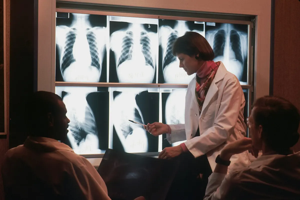
The x-ray technique is now an inseparable part of modern medicine. X-rays are valuable because they can pass through most materials, including human tissue, as long as they aren’t blocked by something dense like metal or concrete. That’s why they’re used to create images like an X-ray taken to diagnose bone and tissue structure inside the human body.
For example, you would have seen x-ray images taken at your dentist’s office. Your teeth appear white because they don’t absorb much X-ray energy, leaving a clear shadow on film when exposed to x-rays from above. Meanwhile, bones appear dark because their high density absorbs most incoming photons before they reach the skin surface, where we see them reflected out into space to record with cameras mounted near our faces inside cabinets explicitly designed for this purpose.
Many diagnoses are conducted with the help of x-ray techniques. For example, a study conducted in the Mazovian Province of Poland found that x-rays were used 7612046 times in a year. This accounts for 1460 examinations for every 1000 people; the numbers show the prevalence of using x-rays in the medical field.
New Developments in X-Ray #
Since Wilhelm Konrad Rontgen discovered the x-ray, many technological developments have occurred. Here are some of the studies done in enhancing x-ray imaging techniques.
Early Clinical Use #
Within the first year of invention, over a thousand papers were published about x-ray technology. In 1986, the first x-ray imaging equipment was installed in Hamburg-Eppendorf, Germany. The production of x-ray machines accelerated after the first installation, and many devices were soon installed worldwide.
The later developments focused on the tubes emitting electrons in the machines. Rontgen used a large inductor, which was large and heavy. Hence, developments were made to reduce the size of the tubes and make them practical to be installed in hospital settings.
Detecting Microcracks #
A recent development in the x-ray technique was finding bone microcracks. Microcracks are not visible using standard x-ray imaging techniques. Hence, the researchers from UMBC and UMB tested a color-based x-ray technique to find microcracks. They used hafnium particles that appear in color and programmed them so that they latch onto the cracks.
New X-Ray Technique for Detecting Explosive Materials #
While there have been many advancements, the most recent and popular one is using Artificial Intelligence (AI) technology with x-rays. AI has long been used to detect patterns in data and for face recognition. The researchers from the University College London used the two technologies to detect explosives and tumors.
According to the paper published in the Nature Communications Journal, the researchers used two proof-of-concept Convolutional Neural Network (CNN) tests. The first test was done using two widely popular CNNs, GoogleNet and Inception ResNet. The first test successfully detected explosive materials, and the second was conducted. The second test was done to examine the efficacy of the first test in challenging environments. The explosives were hidden in electrical items, such as concealed laptops, mobile phones, etc. However, the AI networks and the x-ray techniques could detect the explosives with 100% accurate positive rates, even in the second test.
The Use of the New AI-Aided X-Ray Technique for Detecting Tumors #
The researchers say that while the research was focused on the security field, the new AI-aided x-ray techniques can also have wide applications in medicine, material science, and industrial testing. The technique was focused on determining the microscopic structure difference. Hence, it can be used in medical fields for detecting tumor cells.
Tumor cells are a result of cancer, which is a complex genetic disease. It is also one of the leading causes of death worldwide. According to data from WHO, cancer caused one in six or around 10 million deaths in 2020. While detecting cancer through standard x-ray imaging is possible, it becomes visible only after it progresses.
However, the new x-ray technique can help detect tumors quickly, well before progressing too much. The primary difference between normal and tumor cells is growth. A tumor makes a cell dead. Hence, these cells do not grow, in contrast to normal cells. This creates a small structural difference between the two types of cells. Since the new x-ray technique uses dark-field imaging to detect even the most minor differences, it can easily detect tumors. This can significantly benefit patients as early detection can result in early treatment, which is vital for controlling cancer.
Conclusion #
The x-ray techniques have come a long way. Many developments have already happened, and many kinds of research are ongoing currently. And this is just the beginning. The researchers have already developed a lens that can sense different materials, from plastic compounds and chemicals to drugs. This would help with border security and has other potential applications for detecting disease.
Related reading